What Your Mouth Is Hiding: The Surprising Things a Dental X-Ray Reveals That Your Hygienist Cannot See With the Naked Eye

What Your Mouth Is Hiding: The Surprising Things a Dental X-Ray Reveals That Your Hygienist Cannot See With the Naked Eye

Your teeth look fine. Your gums feel normal. Your last cleaning went smoothly. Here is why that is only part of the story.

The Illusion of a Healthy-Looking Mouth

There is a particular kind of false confidence that comes from looking in the mirror and thinking your teeth look fine. No visible chips. No obvious discoloration. No pain that has been nagging at you. Everything appears, to the untrained eye and even to a trained one working without tools, to be perfectly in order.

And then the X-rays come up on the screen.

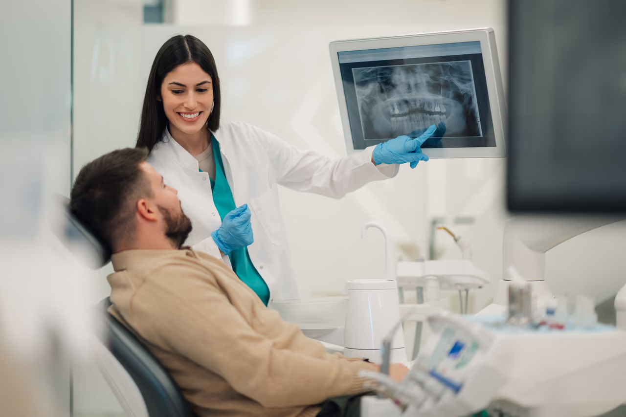
What appears in those images is frequently a different story entirely. A cavity forming in the tight contact between two back teeth, invisible from any angle during a visual exam. A subtle change in the bone level around a tooth that signals early-stage gum disease. A small cyst developing quietly in the jaw, completely asymptomatic, that has been growing for months without the patient having any idea it was there.

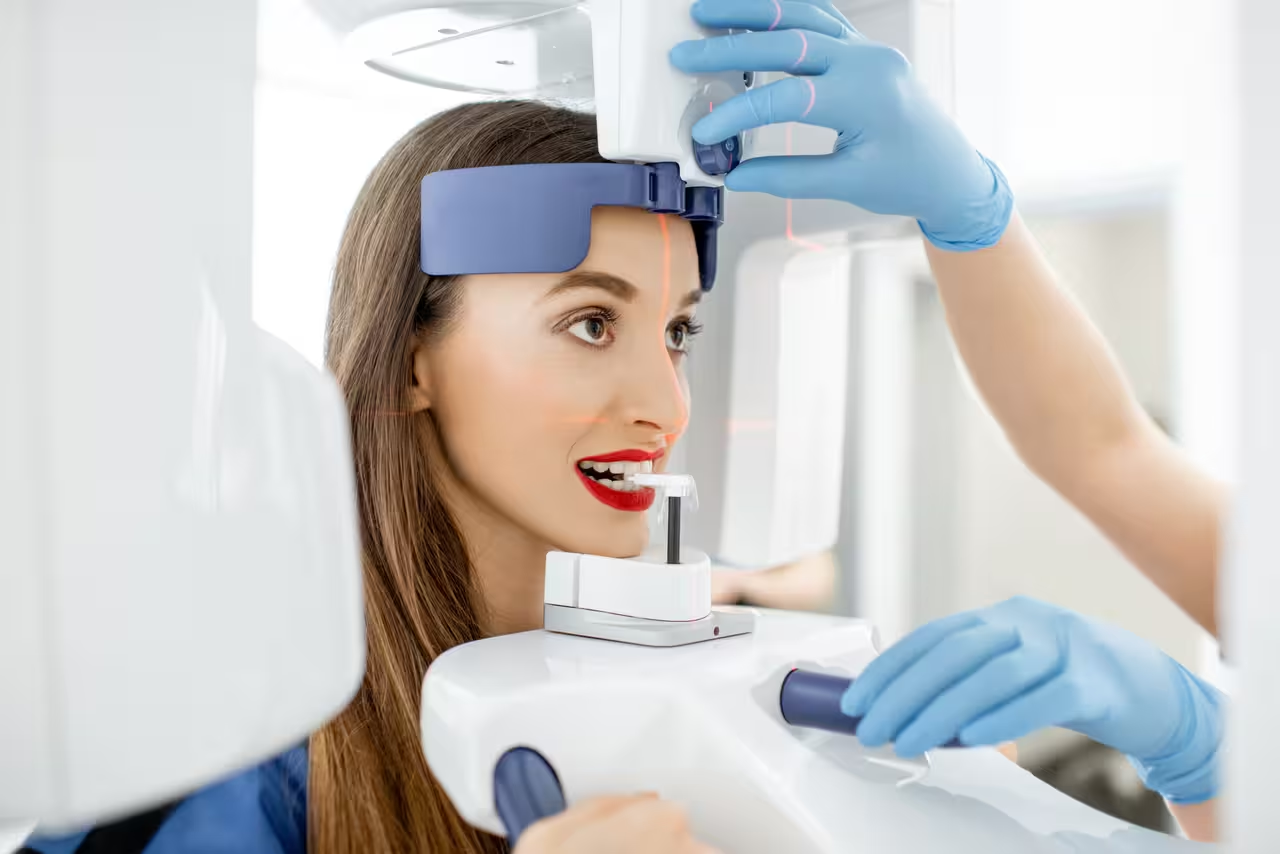
This is not a scare tactic. It is the clinical reality of dentistry, and it is the reason that dental imaging Ewing NJ patients receive at Ewing Dental Associates is treated not as an optional add-on but as a fundamental component of genuinely thorough preventive care.

This post is for every patient who has ever wondered why X-rays are recommended, questioned whether they are really necessary, or simply been curious about what those images are actually showing. The answer is more fascinating and more important than most people realize.

What the Human Eye Can and Cannot See in the Mouth

A skilled dental hygienist conducting a thorough visual examination can observe a remarkable amount of information. The color and texture of the gum tissue. The visible surfaces of the teeth. Signs of wear, staining, and obvious fractures. The general contour of the soft tissues. These observations are clinically valuable and form an essential part of every visit at Ewing Dental Associates.

But the human eye, no matter how experienced the person behind it, cannot see through a tooth. It cannot see between teeth where surfaces are in contact. It cannot see into the bone beneath the gum line. It cannot detect changes in tissue density that have not yet produced visible symptoms. And it cannot see the structures beneath the soft tissues of the cheeks, tongue, and floor of the mouth where serious conditions sometimes develop silently.

Dental X-rays fill this gap because the mouth contains structures that are not accessible through visual assessment. Some of the most significant problems develop first in those hidden areas, even when the clinical examination itself is thorough. 

Seven Things X-Rays Reveal That Cannot Be Seen Any Other Way

Decay Between the Teeth

This is the most common finding on dental X-rays, and it is one of the most instructive examples of why imaging matters. The surfaces where two teeth touch each other, called interproximal surfaces, are completely hidden from visual examination. A cavity forming in that contact area is invisible from outside the mouth and undetectable by probing alone in its early stages.

On an X-ray, early interproximal decay appears as a subtle shadow in the enamel. Caught at this stage, it may be monitored, treated with fluoride, and potentially reversed before a filling becomes necessary. Left undetected, it progresses through the enamel and into the dentin, eventually requiring a restoration or, if it reaches the nerve, more extensive treatment.

Bone Loss From Gum Disease

Periodontal disease, the condition that affects the bone and connective tissue supporting the teeth, is one of the most underdiagnosed conditions in dentistry precisely because its early stages are often painless and visually subtle. The gums may appear relatively healthy while the bone beneath them is already beginning to break down.

X-rays reveal the level and density of the bone surrounding the roots of the teeth with a clarity that no other non-invasive assessment can match. Early bone loss shows up on imaging before it produces the mobility, recession, or sensitivity that patients typically associate with gum disease. Catching it at this stage means treatment is simpler, less invasive, and far more likely to halt the progression effectively.

Cysts and Abnormal Growths

Cysts in the jaw are more common than most patients realize, and they are almost always asymptomatic in their early stages. They develop slowly, often around the roots of teeth or in the spaces where wisdom teeth once were or still reside, and they can grow to a significant size before producing any noticeable symptoms.

On X-rays, cysts appear as well-defined dark areas in the bone, distinct in appearance from the surrounding tissue. Identified early, they are typically straightforward to address. Identified after they have expanded significantly, they may require more involved surgical intervention. This is one of the clearest examples of the clinical value of routine dental imaging: a finding that changes a patient's treatment pathway entirely, discovered during a routine visit, with no symptoms that would otherwise have prompted them to seek care.

Impacted or Poorly Positioned Teeth

Not every tooth erupts the way it should. Wisdom teeth are the most familiar example, but impaction and poor positioning can affect other teeth as well, particularly in younger patients whose dental development is still in progress. An impacted tooth that is pressing against the root of an adjacent tooth can cause resorption of that root over time, damaging a tooth that looks and feels completely normal from the outside.

X-rays reveal the position, angulation, and relationship of all teeth within the jaw, including those that have not erupted and those whose positioning creates risk for neighboring structures. This information guides decisions about monitoring, orthodontic treatment, and extraction that simply cannot be made without imaging.

Root Fractures

A tooth can sustain a fracture of its root that produces no immediately obvious symptoms and is completely invisible during a clinical examination. Root fractures are most commonly caused by trauma, but they can also occur in teeth that have been weakened by previous root canal treatment, large restorations, or grinding habits. The clinical presentation is often vague and may include mild discomfort, occasional sensitivity, or a tooth that simply feels different. These symptoms can easily be attributed to other causes. 

On X-rays, root fractures appear as a break in the continuity of the root structure. Identifying a root fracture changes the treatment plan significantly. A tooth with an undetected root fracture that is restored or crowned without addressing the underlying fracture will fail sooner than anticipated, and often sooner than it would have if the fracture had been identified earlier.

Changes in the Bone That Signal Systemic Conditions

The jawbone is not isolated from the rest of the body. Changes in bone density and structure visible on dental X-rays can sometimes reflect systemic conditions including osteoporosis, certain blood disorders, and metabolic diseases. A dental hygienist or dentist reviewing imaging with an attentive eye may notice a finding that prompts a referral to a physician, even though it has nothing to do with the teeth but may meaningfully affect the patient’s overall health. 

This is one of the reasons that the comprehensive approach to care at Ewing Dental Associates extends beyond cleaning and counting cavities. The mouth is a window into the body, and the information visible in dental imaging is part of that broader picture.

Early Signs That Warrant Oral Cancer Screening Follow-Up

While X-rays are not the primary tool for oral cancer detection, changes visible in the bone and surrounding structures on dental imaging can prompt closer evaluation of the soft tissues. This connects directly to the importance of oral cancer screening Ewing, NJ, patients receive as part of a comprehensive hygiene visit.

Oral cancer screening in Ewing, NJ, at Ewing Dental Associates involves a systematic examination of the soft tissues of the mouth — the tongue, the floor of the mouth, the inside of the cheeks, the lips, the soft palate, and the throat. This visual and tactile examination looks for changes in color, texture, and surface integrity that may indicate early abnormal cellular changes.

The survival rate for oral cancer is dramatically higher when it is detected early. The five-year survival rate for localized oral cancer caught before it has spread is significantly higher than the rate for cancer detected at a later stage. And because early oral cancer is frequently painless and visually subtle, a systematic screening by a trained hygienist at a routine visit is often the only way it gets found at the stage where it is most treatable.

Together, dental imaging in Ewing, NJ, and oral cancer screening in Ewing, NJ, form a comprehensive picture of the patient's oral health that goes far beyond what a cleaning and a visual check alone could ever provide.

The Safety Question: How Much Radiation Are We Actually Talking About?

This is a legitimate question and one that deserves a direct, honest answer.

Modern digital X-rays use a fraction of the radiation associated with traditional film X-rays, typically reducing exposure by seventy to eighty percent. The exposure from a full set of digital dental X-rays is comparable to the amount of background radiation a person receives from their natural environment over the course of a day or two. It’s also significantly less radiation than a short airplane flight.

Dental X-rays are among the lowest-radiation diagnostic imaging procedures in all of medicine. Lead aprons and thyroid collars provide additional protective measure during imaging, providing an extra layer of shielding even for an exposure that is already exceptionally low.

For patients who are pregnant, the team discusses timing and clinical necessity on a case-by-case basis and always prioritizes the patient's comfort and safety in that decision. For all other patients, the risk associated with routine dental imaging is clinically negligible, and the diagnostic value is substantial.

At Ewing Dental Associates, X-rays are not taken on a fixed schedule without clinical justification. The frequency and type of imaging recommended for each patient are based on their individual risk profile, their dental history, and the clinical findings at each visit. Some patients need imaging more frequently. Others can go longer between imaging appointments. The recommendation is always individualized.

What Happens at a Comprehensive Visit at Ewing Dental Associates

Ewing Dental Associates is a hygiene-only practice, which means every single visit is focused entirely on one thing: keeping your mouth as healthy as possible through prevention and early detection. There are no restorative procedures competing for time and attention. No upselling. No pressure.

A comprehensive hygiene visit at Ewing Dental Associates includes a thorough professional cleaning, a clinical examination of the gum tissue and tooth surfaces, dental imaging Ewing, NJ, patients can trust using modern digital technology, and an oral cancer screening in Ewing, NJ, conducted systematically and carefully.

When imaging reveals something worth watching, the team explains it clearly, in plain language, without alarm. When the oral cancer screening identifies a change in the soft tissue that warrants a closer look or a referral, that conversation happens with the same directness and care. And when everything looks healthy, patients leave knowing that they have had a genuinely comprehensive assessment rather than merely a surface-level clean and a rushed visual check.

That is the standard of care Ewing Dental Associates holds itself to at every appointment. And it is the reason patients from Ewing Township, West Trenton, Pennington, Lawrenceville, and Hopewell continue to return.

The Takeaway: What You Cannot See Can Absolutely Hurt You

The mouth is not transparent. The conditions that most significantly affect your long-term oral health, including interproximal decay, bone loss, cysts, root fractures, and early abnormal tissue changes, often produce no pain or visible signs in their early stages. They develop quietly, beneath the surface, in spaces that no clinical examination can access without imaging.

Dental X-rays and oral cancer screening are not bureaucratic requirements or revenue generators. They are the tools that allow a skilled hygiene team to see what is actually happening in your mouth, in its entirety, rather than only the part that is visible to the eye. And the difference between what those tools reveal and what a visual exam alone can find is often the difference between a simple, affordable intervention and a complex, expensive one.

At Ewing Dental Associates, these tools are used thoughtfully, explained clearly, and integrated into every comprehensive visit because the team genuinely believes that thorough preventive care means seeing the whole picture, not just the surface of it.

Ready to schedule your next comprehensive cleaning and checkup? Book online at ewingdental.com or call (609) 771-4111. Located at 177 Scotch Road, Ewing Township, NJ 08628. Next-day appointments available.